Monday, 10 October 2016

The essentials of a scientists gene editing toolbox: CRISPR cas9 system

In my previous post I gave a brief introduction into the CRISPR system. Which effectively is like an immune system adopted by bacteria to protect themselves against viral infections. But still the question is....

How are scientists using this to edit genes in research?

Sit back and relax and for the next part of the CRISPR story to unfold....(crisps not necessary)

Professor Jennifer Doudna and  Emmanuelle Charpentier were studying the bacteria Streptococcus Pyogenes  and discovered the cas9 system (note cas= CRISPR associated proteins). Streptococcus Pyogenes  only have cas9 type cas proteins.


Cas9 Protein (blue) in action with gRNA (cyan) and DNA (magenta)


So, what is the BIG deal about this cas9 protein?

Cas9 protein has a  nuclease region in its major structure-i.e it has an areas within the protein that can cut DNA.  Streptococcus Pyogenes makes two long strips of ribonucleic acid (RNA). RNA is similar to DNA, it is made up of nucloetide bases but unlike DNA RNA is single stranded and contains the nucleobase uracil instead of thymine.

Comparison of DNA and RNA
Cas9 holds both CRISPR RNA (crRNA) which contains a spacer segment which matches up to the corresponding viral RNA. CrRNA is effectively a copy of part of the CRISPR genes (DNA). In my previous post I covered a bit about the spacer segments in the CRISPR system matching up to viral DNA. Cas9 also holds trcrRNA which holds the crRNA in place in the cas9 protein.This allows the viral DNA to be cut by the nuclease activity of the cas9 protein. As the crRNA matches up the viral DNA to be degraded and disposed of and the trcrRNA anchors the crRNA in place. 

Cas9 CRISPR system with viral DNA matching up with crRNA and CRISPR spacer


Scientists exploited the cas9 system by modifying the crRNA by putting their own RNA sequence in to replace the spacer segment and connecting the crRNA and trcrRNA together to form a crRNA-trcrRNA chimera. A chimera is a mythological creature which is made up of different parts of different animals. Similarly in molecular genetics a chimera is a DNA or RNA molecule formed from two or more organisms by laboratory manipulation. This chimera in the cas9 CRISPR system is often referred to the guide RNA or gRNA. 
gRNA and cas9
In short, the cas9  CRISPR system has the cas9 protein which cuts the DNA and the gRNA which guides where the DNA is going to be cut.
An exciting tool for gene editing!

Steps to gene editing using the cas9 CRISPR system....

1) Find and identify the region of DNA that you want to cut
2) Create a guide RNA or gRNA that has a corresponding piece of RNA to the DNA that you want to cut
3) The DNA will be fed through the cas9 CRISPR system and the cas9 protein will cut the selected DNA sequence and inactivate that gene.

Of course the cell will naturally try and repair this break in the DNA by a number of mechanisms causing a mutation. But essentially the selected gene has been inactivated.
Further  from inactivating the gene, a new gene can be inserted.

4) Inserting a host piece of DNA into the cell along with the cas9 CRISPR system so that when the DNA is cut this piece of DNA bridges the cut and the cell repairs and incorporates this new gene into the DNA.

Tada! Genes edited.

Next time...
 In my next post I'll be looking at some of the applications of the cas9 CRISPR system and exploring its future implications. Where will this gene editing take us?

Monday, 26 September 2016

CRISPR, not a new savoury snack but an exciting tool for scientists-an introduction

An introduction to CRISPR in a short series on gene editing

CRISPR is a biological system scientists are exploiting using to edit genes in research. 

I  hear you  saying why, what, where and HOW??? Well grab some CRISPs and hold on tight for this whistle stop tour of CRISPR system!

Firstly, a gene is a short sequence of DNA- deoxyribonucleic acid. Hailed for being the blueprint or the code for life DNA is made up of two strands of four different nucleotides, adenine (A), cytosine (C), guanine (G) and thymine (T) which form a code for the production of proteins in the cell. Every three nucleotides (beads) on one strand form a code for an amino acid.

DNA made up of four nucleotides
In  some diseases there can be changes in this code of nucleotides, for example in cystic fibrosis there is a deletion of three nucleotides which form the code for the the 508th amino acid (phenylalanine) in a protein which lies across the membrane (enclosing) of cells. This is the molecular basis of cystic fibrosis and shows how genetic changes can have a huge impact on cell function. Hence being able to edit this genetic code could have huge implications on health and disease. To explore more about how the amino acid sequence affects proteins have a read of my previous blog post on protein folding and organisation.

What if we could change these errors in the code? How can we ensure that the DNA (which is so long) is edited in exactly the right place? What can we use to do this?

In comes CRISPR (Clustered regulatory inserted short pallindromic repeats-SUCH A mouthful) which was  co-discovered in 2012 by Professor Jennifer Doudna and  Emmanuelle Charpentier who were studying how prokaryotes (i.e bacteria etc) defend themselves against viruses and virus related microorganisms such as phages. Yes, bacteria does have its own form of immune system.


When a bacterium is attacked by a virus, the virus inserts its own genetic material which is then replicated by the bacterium and is used to make new viruses.

When a bacterium has a CRISPR system, it has sequences of identical DNA which are evenly spaced by DNA which is has a unique sequence. This spacer DNA is important as it matches up with viral DNA. 

This CRISPR system is associated with a set of genes called Cas (CRISPR assoicated genes) which encode for proteins that unwind and cut the DNA When a bacterium is infected by a virus and the viral DNA is inserted and the spacer DNA, so the CRISPR gene that matches the inserted viral DNA is copied into a transcript called CRISPR ribonucleic acid (crRNA). The crRNA fits into the Cas protein and this breaks up the viral DNA preventing the infection.


CRISPR system when viral DNA matches CRISPR spacer

When viral DNA is inserted into a bacterium that does not have a matching CRISPR spacer, a different kind of Cas protein is produced that copies this viral DNA to create a CRISPR spacer for the next time it infects as well as breaking down the viral DNA.


CRISPR system when viral DNA does not match CRISPR spacer


That is is the CRISPR system in a crisp packet! Scientists Professor Jennifer Doudna and  Emmanuelle Charpentier studies the CRISPR system in bacteria Streptococcus Pyogenes. This enabled them to develop a tool for gene editing.


Now you know a little bit more about the basic CRISPR system, sit tight for part 2 to explore how this system is used for gene editing and to think about the future implications.


Wednesday, 15 April 2015

Scientists share recipes: communication needed in synthetic biology

The language of scientific research is English: unlike many scientists around the globe I do not have to be at the very least bilingual to pursue this career path. For precision communication is key.

(1) Scientists communicate


How does research move forward? Of course, the sharing of ideas. 

Have you ever had a brilliant idea? For example, a new recipe. You share it with your friend over a cup of coffee. They suggest adding a different ingredient or altering a cooking process based on them coming up with a similar recipe. The combination of ideas  improves the original recipes to produce a dish that is on a whole new level.

However... what if you both spoke different languages? It would make it a difficult task to communicate to technical information. Similarly what if you just gave them the end result (like the MasterChef palate test) and asked them to reproduce the dish?
You would expect different results.

(2) Palate test on MasterChef produces differing dishes

Science does have similarities to cooking. Following a protocol (recipe); using different materials (ingredients) and various equipment (utensils).

In order to move some of the newest areas of science forward, a common language and practices need to be exercised.

The emerging area of synthetic biology ( a cool exciting area!): it combines various areas of biology to create  new biological features, such as: enzymes, biochemical pathways and cells. Effectively engineering but with biological materials. It also involves the redesign of natural biological systems.

Okay, this all sounds interesting... but how does this relate to you? The big dream for synthetic biologists is a future where fuel, drugs, chemicals and food are manufactured by microbes. Yes there is still a long way to go to move from the lab bench to biofactories.

As a scientist it is important to be able to reproduce your results (although if a result is reproducible it doesn't necessarily mean that it is right). So meticulously recording your exact methods and the technical information about your materials and experimental parameters is essential-like sharing a recipe with a friend. You cannot leave out some of the details about the cooking process.

For new emerging fields such as synthetic biology the need for standard ways of reporting technical information is paramount for the field to develop.

After all, what we can achieve together is far greater than what we can achieve alone.

References

Hayden E, 2015., Synthetic biologists seek standards for nascent field., Nature news, 520, pp 141-142, doi: 10.1038/520141a

Figures


Monday, 17 November 2014

The cell's ironing: protein folding and folding surveillance

Proteins are highly organised structures: they're folded into particular conformations;associated with other protein subunits and have different levels of organisation (see previous post).
But how do they achieve their folded state?
(1) How do proteins fold?
We all like help-so do proteins
Sometimes it is easier when someone helps you fold away your clothes. Same for proteins-yes they are largely self-folding. But what happens when this goes wrong?

Chaperones are part of the cell’s emergency services attending various accidents (misfolded proteins) but in a disaster they all flood to the scene (when a cell is under stress).Many chaperones are heat shock proteins (Hsp) and are highly abundant when a cell is under stress (during nutrient starvation, extreme temperatures, exposure to toxins etc).

Aggregates can easily form when hydrophobic regions of the protein can end up on the outer surface of the misfolded or partially folded protein and will attract other misfolded proteins with exposed hydrophobic residues (as the cell has an aqueous environment the water fearing residues want to bury themselves in the core of a protein).  In a living cell it not just empty space (imagine trying to fold your washing in a crowd without elbowing anyone!). Chaperones help the protein fold in the most energetically favorable way as well as preventing the unwanted binding of other proteins during the folding process.

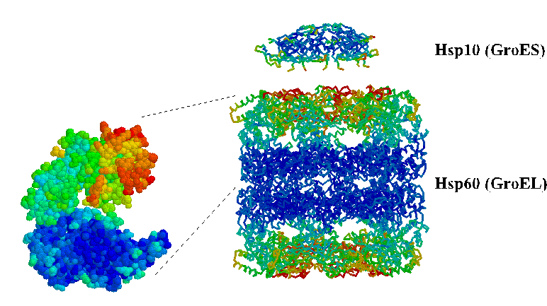
Some molecular chaperones (Chaperonins-Hsp60) act like a huge tent in the middle of the crowd where you can fold your washing without people getting in the way. They completely isolate the protein from the environment of the cytosol.

(2)Bacterial  Hsp60 (GroEL) and associates Hsp10 (GroES)


Whilst other chaperones (Hsp70) bind to a region of the protein to aid folding like having a group of people who are designated to aid in folding of washing-it is their job.

(3) Hsp70 binds to hydrophobic regions to aid folding


Chaperones are not just involved in folding they’re also important for unfolding, refolding and trafficking.  Chaperones work by an ATP* dependent mechanism: they bind to hydrophobic regions of a misfolded (non-native) protein.

One size of chaperone doesn't fit all:
There are different classes of molecular chaperones (they are usually classified by their molecular weight).  Hsp60 (chaperonins) ,Hsp70,Hsp90 and Hsp100 systems. The numbers indicate the molecular mass of each Hsp protein subunit.

Focusing on the chaperones involved in protein folding:  namely- Hsp60,70 and 90.
They generally act under two systems Hsp70 and Hsp60 (chaperonins).

Hsp70 interacts with newly synthesised proteins and releases them . Whilst chaperonins  (Hsp60) are large cylindrical protein complexes ( figure 3) (composed of several polypeptide chains-quarternary structure: an outfit (see previous post)).They enclose the proteins for folding. The two systems act sequentially Hsp70 act on newly synthesised proteins and then chaperonins assist folding of proteins that do not reach their correct folded state by Hsp70 alone.

So imagine you (Hsp70)are folding your washing and your Mum (Hsp60) is stood by your side and when something is difficult to fold or you have not folded it correctly your Mum takes over to ensure correct folding.

The Hsp90 system functions downstream of Hsp60. It acts in the late folding stages of proteins involved in cellular signalling and development. Both Hsp70 and Hsp90 can direct proteins for degradation.

It is now like your Grandma (Hsp90) has joined the folding party; smoothing out the creases of odd item that your Mum (Hsp60) has missed.

Folding avoids creases but what does it mean for proteins?

When proteins fold they gain a particular shape which is important for the function of that protein. It may allow it to associate with other proteins to form a complex.

It has been suggested that the decline in the cell’s ability to correctly fold proteins and maintain correct protein folding (proteostasis) declines with age. This can allow the formation of protein aggregates which are involved in many neurodegenerative diseases such as Alzheimer’s and Parkinsons disease. Finding out more about chaperone mediated surveillance of proteins is vital as these diseases become more prevalent in our aging society.
(4) Comparison of a healthy neuron with a Alzheimer's neuron with protein aggregates ( Beta amyloid plaques and neurofibrillary tangles)
*ATP= Adenosine triphosphate is the energy currency of the cell. It is a phosphorylated nucleotide that is made and consumed by all cells and drives chemical reactions

References

Hartl F, Bracher A, Hayer-Hartl M., 2011. Molecular chaperones in protein folding and proteostasis., Nature reviews., 475., pp324-332

Lodish H et. Al., 2008., 6th Ed., Molecular cell biology., W.H.Freeman., New York

Saibil H., 2013. Chaperone machines for protein folding, unfolding and disaggregation., Nature reviews., 14., pp631-632

Images


Tuesday, 28 October 2014

If proteins were in my wardrobe: folding and organisation

So my blog has been very quiet (apologies). A new start to the academic year so a new blog post!


When you open your wardrobe, how does it look?
Like this?
(1) Clean and tidy


...Or admittedly more like this?
(2) Disorganized and messy


Many things in molecular cell biology can appear rather chaotic on the surface. Proteins look like highly higgledy piggledy molecules. When  actually they’re pretty organised things: structurally complex and sophisticated molecules of life.

How the cell folds its wardrobe


Amino acid sequence: The shape of a protein is determined by its amino acid sequence (primary structure)- the threads of the garments (20 types in total). They can be grouped into different categories according to their side chains-acidic, basic, uncharged polar and nonpolar.

(3) 20 Amino acids 


Non covalent bonds also influence the folding of proteins: hydrogen bonds, ionic bonds and Van der Waals attractions.

Environment: The cell has a very aqueous environment which affects a proteins shape, making it very compact. The non polar side chains are hydrophobic (water-fearing) bury themselves in the core of the proteins whilst the polar (hydrophilic- water-loving) side chains interact with water and so tend to gather on the outside of the protein.

When completing a task we often look for the easiest way to accomplish the task with minimal cost (whatever that may be) Proteins do the same: they fold into a shape of the lowest energy (i.e energetically favourable to maintain- the easiest way to fold).

Organisation of protein structure

Unlike my wardrobe proteins have a very organised structure that can be broken down into four categories:
(4) Categories of protein structure 

Many proteins have large structures (of one very long string on amino acids folded up) which gives a further unit of organisation: the protein domain.

 This is a length of the protein which is between 40 and 350 amino acids (stitches) in length. This length folds independently to the other regions of the protein like different parts of a shirt (collar, sleeve,  the pocket) are all still part of one shirt but and useless on their own yet still independent.
Different protein domains often are associated with different functions.

Items in a cells wardrobe (some examples)


Src protein kinase:  One of the many secretaries of the cell. It acts in signalling cascades by adding phosphate groups from high energy donor molecules like ATP to substrate molecules.
A protein formed of four different domains (each has a different function).

The SH2 and SH3 domains are involved in regulation whilst the catalytic domain and the activation loop play a role in the catalytic activity of the kinase.

(5) Src Kinase Hck


Haemoglobin: Found in red blood cells and vital for the transport of oxygen around the body and also has roles in the transport of carbon dioxide and hydrogen ions. It is a multi-subunit protein: two α subunits (two protein chains of 141 amino acids-stitches) and two β subunit (two protein chains of 146 amino acids-stitches).

(6) Haemoglobin
So next time you open your wardrobe to put your clean clothes away (freshly synthesized (nascent) proteins) think about how organised our cells are: folding proteins and packaging them to be sent to various locations within the cell. How organised is your wardrobe?

Upcoming posts
More about protein organisation and folding-why and the cellular machinery involved
What happens when folding goes wrong?

References
Alberts B et al., 2008., 5th Ed., Molecular biology of the cell., New York., Garland Science
Berg  J et al., 2011., 7th Ed., Biochemistry., New York, W. H. Freeman

Images


Tuesday, 28 January 2014

I'm sorry, bio-what??!!-A brief plug for biochemistry

University is a time where you meet lots of new people. Being in a new city and getting involved in a new community. You get asked the three questions a zillion times:
1)What's your name?
2)Where are you from?
3)What are you studying?


A tip for you all!
 
Okay so I study biochemistry. But I thought I'd give an insight into how diverse a subject it is.

Biochemistry is not quite a fusion of your biology and chemistry lessons you might've had at school
It is much more than that!

Biochemistry is at the heart of many areas of the life sciences such as genetics, cell biology, energy and metabolism, plant biology and development of disease.

Lubert Stryer, the famous biochemist and author of Biochemistry (W.H. Freeman & Co.), states that biochemistry is “rapidly progressing from a science performed almost entirely at the laboratory bench to one that may be explored through computers. Its practical approach applies the molecular aspects of chemistry to the vast variety of biological systems."

 The thing I love about studying biochemistry is taking a 'simple' cell and finding out the intricate mechanisms that enable that cell to function. For example protein synthesis: DNA is transcribed into messenger RNA (ribonucleic acid, a molecule similar to a single strand of DNA). The code of bases that the mRNA contains is transcribed by ribosomes into a chain of amino acids joined by peptide bonds a.k.a a protein.

That may sound simple summarised in just a few short sentences.But it's far from it!  And that is only one example!

Molecular biology shows that there is to life that meets the eye which makes me feel privileged to be studying biochemistry.

Upcoming posts
Sugar and sweetners-What's the difference?

Saturday, 30 November 2013

The gut feeling about caffeine

 Following from my previous caffeine post: What are the effects of caffeine on our bodies? I thought it's time to round off my investigation into the intriguing substance of caffeine.

Not the best way to find out the effects of caffeine on the body


Do you ever get that gut feeling when drinking coffee?-Coffee and the gastrointestinal system

There is strong evidence that coffee increases gastric acid secretion. Interestingly experiments have shown that it's the other constituents in coffee that contribute to this increase in gastric acid secretion.
This was shown by  measuring the dose response of caffeine, regular coffee and decaffeinated coffee for gastric acid secretion in normal subjects. Both regular coffee and decaffeinated coffee gave a similar response in gastric acid secretion which was higher than that of caffeine alone (on a cup equivalent basis) (Cohen and Booth, 1975).

The dose response? what??

Measuring the dose-response enables scientists to observe the change in an effect (in this case:gastric acid secretion) caused by varying the levels of dose of a substance (in this case: dosage of caffeine, regular coffee and decaffeinated) after a certain length of time.

Kidney function

The kidneys play an important role in filtering the blood. The kidneys remove waste products (such as urea) and extra water from the blood which form urine. Any drinker of caffeine beverages is well aware that caffeine tends to stimulate an increased flow of urine.
Anatomy of Kidney

Caffeine was traditionally used to increase urine output until more potent diuretics became available. The diuretic effects of caffeine appear to be due to an increased rate in blood flow to the kidneys and increased rate of blood filtration. These affects are due to antagonism of circulating adenosine (see previous post) having a regulatory role in the formation of urine  (Fredholm 1984).

So will my cup of coffee cause a fluid imbalance?
A dose of 300mg of caffeine (approximately 4-5 cups) can cause acute diuresis- this has been shown by several studies (Oswald and Schnermann., 2011). Caffeine will only cause a significant increases in the volume of urine excretion and a negative fluid imbalance in a large dose. A study in which caffeine was given (6mg/kg) for 11 days showed no effect in daily urine volume (Armstrong et.al 2005).

Time for a breather-Respiratory system

Caffeine is a respiratory stimulant (Braun 1996). However based on the average person's caffeine consumption has little effect on the respiratory system. Larger doses of caffeine has proven to be effective in the treatment of neonatal apnea-the cessation of breathing in newborns.

Upcoming posts

How about  I keep it a surprise this time?


References

Armstrong LE, Pumerantz AC, Roti MW, Judelson DA, Watson G, Dias JC, Sokmen B, Casa DJ, Maresh CM, Lieberman H, Kellogg M. 2005., Fluid, electrolyte, and renal indices of hydration during 11 days of controlled caffeine consumption. Int J Nutr Exerc Metab. 15

Braun S., 1996., Buzz: The Science and Lore of Alcohol and Caffeine., Cary., NC., USA., Oxford University Press
., 252-265. Cohen, S., and Booth, G. H. 1975., Gastric acid secretion and lower-esophageal-sphincter pressure in response to coffee and caffeine., New England Journal of Medicine., 293,897-899.

Fredholm, B. B., 1984.,Cardiovascular and renal actions of methylxanthines., New York:Alan R.Liss

Oswald H and Schnermann J., 2011., Methylxanthines and the Kidney., Handbook of experimental pharmacology., 200., 391-412.

Monday, 25 November 2013

Why can't the traffic on our roads be more like the traffic in cells?

Time to take a short break from caffeine. After all it's a special occasion with the announcement of the Nobel Prize 2013 winners! (It's about time I did a post about it)

I see science being a bit like a mystery to be solved: all the clues have to be gathered together to see the overall picture. However like scooby doo and his gang it is much easier solving the mystery together!

Likewise this years noble prize in medicine was awarded to three scientists: Randy Schekman, James Rothman and Thomas Südhof. Who uncovered how the cell organizes the transport of substances around the cell.
Cells have traffic?

So you're probably thinking how do cells have traffic?

Rules of the road: Cell style

Substances are transported around the cell in membrane bubbles called vesicles (these are a bit like taxis transporting passengers from A to B). Vesicles bud and pinch-off from specialised structures in cells called organelles. Vesicles travel along the cytoskeleton of the cell like taxis travel along roads. The cytoskeleton of the cell is a network of protein filaments and tubules in the cytoplasm of many cells. As well as its role in transport the cytoskeleton gives the cell shape.

Red showing cells actin cytoskeleton


Randy Schekman's contribution

He discovered a set of genes that were required for vesicle transport.
How did he do this?
He studied the genetic basis of how a cell organises its transport system by using yeast as a model. (Yeast  are often used experimentally as they can be grown quickly and have a eukaryotic cell structure-they have membrane bound organelles like human cells, we also share many genes with yeast cells). Sometimes the best way to discover how something works is to look at what happens when that system goes wrong. That's exactly what Schekman did. He studied yeast with defective transport systems, identified the cause of congestions and identified the mutated genes. So he had solved one aspect of cellular transport.

James Rothman's contribution

He revealed the proteins involved in the fusing of vesicles (the transporters of cellular cargo)  with their targets (their desired destination in the cell) enabling the vesicles to deliver their cargo. Through his experiments he showed that these proteins bind in specific combinations to ensure that cargo is delivered to a specific location.

Thomas Südhof

He was interested in how nerve cells communicate with each other in the brain. He showed how vesicular contents can be released as a result of a cell signal.

In nerve cells signals come in the form of neurotransmitters (chemicals such as acetylcholine and noradrenaline). Nerve signals pass from nerve cell to nerve cell at junctions called synapses. Südhof found how nerve cells release neurotransmitter into the synaptic cleft upon a signal.

When an action potential (a electrical signal) arrives at a nerve terminal, calcium ions enter the cell through a channel that is temporarily open due to the arrival of the action potential. The entry of calcium  causes vesicles containing neurotransmitters to be released into the synaptic cleft where the signal is communicated to the neighbouring nerve cell. Südhof discovered the calcium sensing proteins in the pre-synaptic nerve terminal which direct and dock vesicles containing neurotransmitter to the cell membrane where the neurotransmitter is released into the synapse (using the mechanisms discovered by Schekman and Rothman). See home-made diagram below (apologies I am no artist).
Synapse- Signalling between two nerve cells

Cells are incredible at transporting substances. It's interesting to see how the work of a few scientists have uncovered the mechanism of transport of substances. Hats off to them! I hope next time you're travelling you'll think about the trillions of cells in your body transporting substances to and fro.

Upcoming posts
More posts on caffeine are inevitable. Will hopefully be looking into reviewing scientific papers in the future-Watch this space.

Thursday, 3 October 2013

What are the effects of caffeine on our bodies?

An advertisement in a London newspaper in 1657 described coffee as: "A very wholesome and physical drink that helpeth digestion, quickeneth the spirits and maketh the heart lightsom, is good against eye-sores,coughs, colds, rhumes, dropsie, gout and scurvy." (Braun S., 1996)

Obviously today we do not view coffee and caffeinated substances to be this marvellous medical elixir.

So what are the effects of caffeine on the different systems in our bodies?

I'll be starting with the nervous and cardiovascular systems
Nervous and cardiovascular systems
Caffeine and the nervous system

Caffeine is a central nervous system stimulant-It increases brain and spinal cord activity, speeding up physical and mental processes.

Caffeine also affects the autonomic nervous system -A branch of the peripheral nervous system which controls smooth muscle, internal organs and glands (click for more information on the nervous system).
There has been reports that caffeine increases skin conductance (sweat gland activity). A 500 mg anhydrous (dry weight) caffeine dose can produce behaviour similar to General Anxiety Disorder also referred to as 'caffeinism' (Bruce et al., 1986). Caffeinism is a condition resulting from ingestion of a large dose of caffeine shown by diarrhoea, elevated blood pressure, restlessness and insomnia.

Putting things into perspective: a 500 mg dose all at once is a large amount considering  an average cup of instant (heaped tea spoon) coffee contains 60 mg (Center for science in the public interest., 2012). A large dosage of caffeine can produce extreme symptoms due to the rapid rate at which  caffeine is absorbed. So do not fear drinking your morning cup of coffee as 500 mg is a high dose and was used for experimental purposes.

Note how some of the effects caffeine has on the nervous system appear anxiety reducing (i.e increasing concentration and reducing tiredness) and some effects similar to anxiety (insomnia and restlessness).

Cardiovascular system

 Caffeine has an interesting effect on blood flow. In moderate amounts caffeine can increase blood pressure-the effect is more significant in those who do not regularly consume caffeine (Bruce., 1986: James 1997). But surprisingly has little effect on heart rate (France and Ditto 1992).

How does this change in blood flow come about?
Caffeine molecules compete with adenosine for adensosine receptors on cardiovascular cells (See previous post on receptors). In the cardiovascular system adenosine causes dilatation of coronary and cerebral blood vessels. Caffeine inhibits this process, meaning the blood vessels remain constricted. The narrower diameter of the blood vessels result in higher blood pressure (James 1997).

Time for a little analogy......
Imagine you have gone shopping for a new garden hose. Your standing in the aisle looking at the different diameters of garden hoses you can buy. Whilst you do this you imagine watering your garden with your new hose. The pressure of the stream of water coming out of the end of the hose depends on the diameter of the hose. Logically the narrower the diameter of hose the higher the pressure.

Hoses varying in diameter
Like hoses our blood vessels are the same. The narrower the vessel the higher the blood pressure in that vessel. But blood vessels (unlike your garden hose) are clever and can change diameter.

When adenosine is present in the blood, this causes the smooth muscle in the blood vessels to relax- the vessel wall gets thinner and the space where blood flows in the vessel gets wider: this is vasodilatation. When caffeine is present and binds to adenosine receptors instead of adenosine; it has the opposite effect. The smooth muscle in the vessel wall contracts and gets thicker so the space where blood flows gets narrower resulting in a higher pressure in that vessel: this is vasoconstriction.

Upcoming posts....

I'll be looking at the physiological effects of caffeine on the gastrointestinal and respiratory systems and also the effect on renal function (think peeing!). I also hope to do a bit about caffeine and health.

References

Braun S., 1996., Buzz: The Science and Lore of Alcohol and Caffeine., Cary., NC., USA., Oxford University Press

Bruce M, Scott N, Lader M, Marks V., 1986., The psychopharmacoloical and electrophysiological effects of a single dose of caffeine in healthy human subjects., British Journal of Clinical Pharmacology., 22, 81-87., [Online]., Available at: http://www.ncbi.nlm.nih.gov/pmc/articles/PMC1401080/?page=1 ., [Accessed: 02/10/2013].

Center for science in the public interest., 2012., Caffeine content of food & drugs (online)., (Updated 2012)., Available at: http://www.cspinet.org/new/cafchart.htm., [Accessed: 03/10/2013].

France C and Ditto., 1992., cardiovascular reponses to the combination of caffeine and mental arithmetic, cold pressor and static exercise stressors., Phychophysiology., 29., 272-282.

James J., 1997., Understanding Caffeine., California., USA., SAGE Publications Inc

Friday, 27 September 2013

Puzzling over caffeine and adenosine-it's time to talk about receptors (please don't be antagonistic!)

Heading back to university for the start of the academic year it'll be inevitable that my coffee consumption will increase exponentially. So I'm back again and thinking about what happens in my body when I consume my daily caffeine fix.

After looking at caffeine metabolism and whether caffeine is addictive and causes insomnia in my previous posts. I thought it was time to start looking at HOW caffeine changes our body's functioning.

So time to start fitting the pieces together of the physiological effects of caffeine. This starts with receptors.

What are receptors?

Receptors are proteins which can be found on cell membranes. They bind to specific chemicals (these could be hormones, drugs, neurotransmitters etc). The binding of these chemicals can cause a change to occur in the cell.

It is natural when looking at the biological effect of caffeine in the body to start looking at what happens when caffeine binds to a receptor.

The caffeine-adenosine relationship

Everybody has two friends which are uncannily similar to each other. Caffeine and adenosine are those uncannily similar friends-they have a similar molecular structure.


Spot the difference for the chemists!
These similarities in structure mean that caffeine can bind to adenosine receptors and block their function. Adenosine on binding to its receptor generally inhibits physiological activity (it makes us feel more tired). Due to adenosine and its half brother caffeine having a similar molecular structure caffeine blocks adenosine receptors (mainly  A1 and A2 types of adenosine receptors) preventing their activation resulting in caffeine having a stimulant effect. Hence caffeine is an antagonist (It interferes with the binding and physiological affect of adenosine).


Still puzzled?

So if that’s perhaps gone over your head a little......

 Imagine you are putting together pieces of a puzzle. You are looking for a piece of the puzzle that joins to the piece you have in your hand (the adenosine receptor). You can see two similar looking pieces on the floor (an adenosine and caffeine molecule) that look like they'll both join onto the piece you are holding. You try piece A (the caffeine molecule) and find that it pretty much fits onto the piece you are holding (the receptor) but the fit is not perfect. Despite this you decide to continue putting the puzzle together. However you find that you cannot fit the rest of the puzzle together and complete it (receptor has not been activated so the physiological effect is inhibited-completing the puzzle) as the two pieces you first joined together are not the correct pieces to join to complete the puzzle (the physiological effect is inhibited).

You go back to the original piece you had in your hand (receptor) and this time you join piece B (the adenosine molecule) onto it and find that these two pieces fit perfectly together. This time you find you are easily able to complete the puzzle (the receptor is activated resulting in the full physiological effect-the puzzle is completed) as all the other pieces fit around the two you first joined together (the receptor and adenosine molecule).

Let’s put the puzzle together....

The binding of caffeine to adenosine receptors prevents adenosine from binding to the receptor causing a change in the cell's behaviour. Caffeine inhibits the effect of adenosine. This is how caffeine can make us feel more awake. As adenosine decreases neurotransmitter release, dilates blood vessel and inhibits lipolysis (break down of fats)-these effects make us feel more tired. Caffeine preventing this in turn makes us feel more awake.

Upcoming posts....

Caffeine and its effect on the physiological processes in our body- how this stimulant takes its effect on the nervous, gastrointestinal, respiratory systems and renal function.

Sunday, 1 September 2013

Lets try and resolve some caffeine confusion


As I settled down to read the newspaper with (yes) a cup of coffee! An article about coffee caught my eye. "More than four cups of coffee a day increases risk of early death" (check it out and leave your comments).

Again the mysterious cup of coffee causes a stir. 

Drinking coffee is such a prominent part of the modern lifestyle with coffee shops clustering in every high street and our supermarket shelves stacked with all kind of coffee. It surprises me how variant
peoples views are on the effects of this seemingly everyday essential to our health is.


Caffeine is mainly found in coffee, tea, energy drinks, chocolate, cold and flu medicines and surprisingly some caffeine is still found in a cup of decaffeinated coffee (approximately 3.5% of the caffeine found in an non-decaffeinated coffee of the same strength and volume).


Sources of caffeine

Can caffeine be addictive?

Firstly I must define what I am referring to as an 'addictive' substance: a substance which an individual becomes physically and psychologically dependant and has the compulsion to increase the dose of said substance.
Scientists don't class caffeine as an abusive addictive drug. Regular caffeine users may experience a mild physical dependence. Withdrawal symptoms include: headache, fatigue and lack of concentration. However these symptoms may only last a day or so. These symptoms can be reduced is caffeine intake is reduced gradually.
But this still hasn't answered my question as to whether caffeine is addictive?
Well caffeine does not cause severe withdrawal symptoms or the user to exhibit compulsive behaviours as with other drugs. So caffeine is generally not considered addictive, certainly  medically speaking and according to my definition of 'addictive substance'.

Does caffeine cause insomnia? 

It's all about timings....
As mentioned in my previous post caffeine is rapidly absorbed and processed by the body (in about 45 minutes it is distributed to tissues in the body). It takes about 5-7 hours for half of the caffeine to be eliminated from the body (based on dosage from an average cup of coffee). It takes up to 10 hours for 3/4 of caffeine to be eliminated.

So what has this timing got to do with insomnia?
Caffeine is a stimulant so it keeps parts of your brain overactive preventing you from sleeping .
This means that a cup of coffee or two in the morning is unlikely to affect the average persons sleeping habits or consuming caffeine 6 hours before going to bed. However this depends of the individuals sensitivity to caffeine. People who are more sensitive to caffeine may experience insomnia, nervousness and gastrointestinal upset.

Caffeine is known to be a diuretic (it makes us need to pee), consuming a product containing caffeine late at night can mean we won't sleep properly as we keep needing the toilet.

What about increased risk of death?

Caffeine is just a small part of our diet and certainly in small doses it won't impact our health (although a large dose would mean drinking about 100 cups of coffee in a short space of time). Excessive coffee drinking is often linked to other unhealthy habits which may account for the statistics displayed in the article. But remain calm. You're not going to drop dead from drinking another cup of coffee.

Thanks for reading!


Next posts?

I'd like to investigate more of the biological effects of caffeine. And start asking how and why :)
Why does coffee make us pee? How does it mask symptoms of tiredness?